A research team led by Professor Chulhong Kim (CiTE, ME), and Park Byul-lee (CiTE M.S./Ph.D. candidate, advisor Prof. Chulhong Kim) conducted joint research with a research team led by Prof. Yumiao Zhang of the Chemical Engineering Department in Tianjin University to create photoacoustic imaging technology for deep-tissue images. This research was published in Small, one of the top multidisciplinary journals covering nano and microscale science.
Optical Imaging prior to this research could look into less than one millimeter deep but the photoacoustic imaging technology enables us to observe a few centimeters into body tissues. The research team’s imaging technology reduces light scattering and low background signals from biological structures. Their technology first encapsulates the thiadiazoloquinoxaline-based semiconducting polymer (SP) then removes excess surfactant. This is followed with the production of a dual functional polymer system named surfactant-stripped semiconductor polymeric micelles (SSS-micelles), which enables photoacoustic imaging at both 1,064 and 1,300 nm wavelengths. Their technology allowed them to successfully observe the stomach and bladder of rats, which is about 5.8 cm into their bodies. This penetration length has been recorded as the deepest imaging produced as of today.
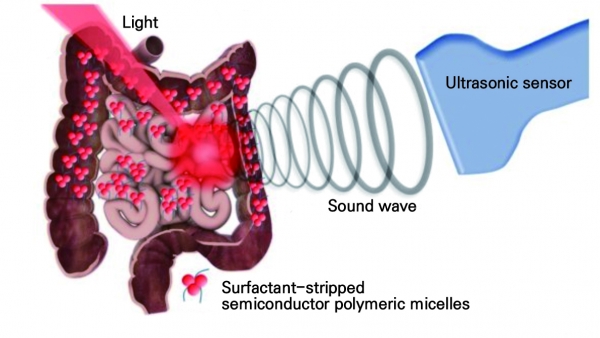
The principle of photoacoustic imaging is the use of ultrasonic sensors to detect the sound waves that are produced when tissues expand after absorbing light. The ultrasonic sensors then use the detected sound waves to create images of the tissues. Until now, most lights used for photoacoustic imaging had wavelengths of 650-900 nm, which is hard to detect deep inside the body. The use of SSS-micelles enabled the detection of lights with wavelengths of 1,064 nm and 1,300 nm.
This newly developed technology has its advantages over currently used CT scans. This new photoacoustic imaging technology does not use X-rays which eliminates radiation exposure dangers. This technology also is used with 1064 nm lasers which are relatively cheaper and can be used commercially. Prof. Kim stated that, “I hope this project continues to achieve good results when developed into clinical research.”
